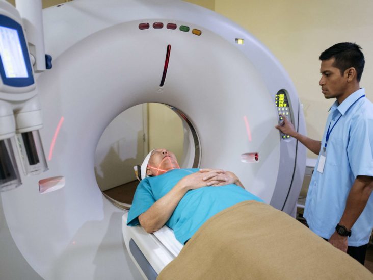
An MRI (magnetic resonance imaging) examination is a painless examination that yields very clear pictures of the structures and edifices within your body. MRI uses a large lodestone, radio surfs and a computer to crop these inclusive pictures.
Because MRI doesn’t use X-rays or other radioactivity, it’s the imaging examination of choice when persons will need recurrent imaging for analysis or therapy monitoring, particularly of their brain.
What is an open MRI?
An open (or “open bore”) MRI denotes the kind of machine made by the MRI Machine Manufacturers that take the pictures. Characteristically, an open MRI apparatus has two level lodestones located over and underneath you with a huge space between them for you to lie. This permits for open space on two flanks and eases much of the claustrophobia many people know with closed-bore MRI apparatuses.
Though, open MRIs don’t take as clear pictures as closed-bore MRI apparatuses. Closed-bore MRI apparatuses have a circle of lodestones that forms an exposed hole or pipe in the center where you’d lie to get the pictures. Closed-bore MRIs are slender with tight head-to-ceiling space. This can reason nervousness and uneasiness for some people, but these MRI apparatuses take the best quality pictures.
If you’re anxious about your MRI examination or have a dread of shut spaces, speak to your healthcare supplier. If required, your supplier will make deliberate choices for tranquilizers (drugs to make you feel tranquil) or even anesthesia is essential.
What is an MRI with divergence?
Some MRI examinations use an inoculation of divergence material. The divergence agent covers gadolinium, which is a sporadic earth metal. When this material is present in your body, it modifies the magnetic possessions of nearby water particles, which improves the excellence of the pictures. This recovers the sensitivity and specificity of the investigative pictures.
Divergence material improves the discernibility of the following:
- Growths.
- Irritation.
- Contagion.
- Bloodstock to certain tissues.
If your MRI needs a divergence material, a healthcare supplier will implant an arterial catheter into a vein in your hand or limb.
What’s the variance between an MRI examination and a CT examination?
Magnetic resonance imaging (MRI) uses lodestones, radio surfs and a computer to generate pictures of the inside of your form, while computed tomography (CT) uses X-rays and computers.
Healthcare suppliers often favor using MRI images instead of CT images to look at the non-bony shares or soft matters inside your form. MRI scans are also harmless since they don’t use the harmful ionizing radioactivity of X-rays.
MRI examinations also take much stronger images of your brain, spinal cord, nerves, muscles, sinews, and ligaments than unvarying X-rays and CT examinations.
Though, not everybody can endure an MRI. The captivating field of MRI can relocate metal grafts or disturb the purpose of expedients such as pacemakers and insulin pumps. If this is the circumstance, a CT image is the next best choice.
MRI skimming is typically costlier than X-ray imaging or CT imaging.
When would I want an MRI?
Healthcare suppliers use magnetic resonance imaging (MRI) to aid identify or screen the treatment for many diverse circumstances. There are also diverse kinds of MRI machines supplied by the MRI Machine Suppliers founded on which part of your form your supplier wants to inspect.
Suppliers use cardiac (heart) MRIs for numerous motives, including:
- To assess the structure and purpose of your heart cavities, heart valves, the scope of and blood movement through major vessels and the nearby structures.
- To identify circulatory circumstances, such as cancers, contagions, and seditious conditions.
- To assess the properties of coronary artery illness, such as restricted blood movement to your heart muscle and blemishing inside your heart muscle after a heart attack.
- To assess the structure and purpose of the heart and blood vessels in broods and grownups with hereditary heart illness.
Form MRIs can assess edifices and identify several circumstances, including:
- Lumps in your torso, stomach, or pelvis.
- Liver illnesses, such as cirrhosis, and problems with your bile vessels and pancreas.
- Demagogic bowel illnesses, such as Crohn’s illness and ulcerative colitis.
- Deformities of blood vessels and irritation of the vessels (vasculitis).
- A mounting infant in the womb.
MRIs of frames and junctions can help assess:
- Bone contagions (osteomyelitis).
- Bone growths.
- Disk irregularities in your backbone.
- Joint problems produced by wounds.
Healthcare suppliers occasionally use breast MRIs with mammography to notice breast tumors, particularly in people who have thick breast tissue or who may be at high danger of breast cancer.
Are MRI examinations safe?
An MRI examination is usually safe and postures almost no danger to the typical person when suitable security rules are tracked.
The robust magnetic arena the MRI apparatuses bought from the MRI Machine Dealers produce is not damaging to you, but it may reason entrenched medical devices to blip or garble the pictures.
There’s a very small danger of an adverse reaction if your MRI needs the use of divergence material. These responses are typically slight and manageable by medicine. If you have an adverse reaction, a healthcare supplier will be obtainable for instant help.
Healthcare suppliers normally don’t complete gadolinium contrast-enhanced MRIs on expectant people due to unidentified dangers to the evolving baby except it’s unconditionally essential.
Who does the examination?
A radiologist or a radiology technician will complete your MRI. A radiologist is a medical clinician who achieves and understands imaging examinations to identify circumstances. A radiology technician is a healthcare supplier who’s specifically trained and specialized to complete an MRI examination.
How does the MRI occur?
Magnetic resonance imaging (MRI) efforts by passing an electric current through looped wires to generate a provisional magnetic field in your form. A spreader/headset in the appliance then directs and obtains radio surfs. The computer then uses these motions to make digital pictures of the skimmed part of your body.
What do I require to do to concoct an MRI?
The magnetic resonance imaging (MRI) scanner uses robust magnets and radio wave signs that can cause warming or conceivable undertaking of some metal substances in your body. This could result in fitness and security issues. It could also cause some entrenched electronic medical devices to break down.
If you have metal-containing substances or entrenched medical devices in your form, your healthcare supplier wishes to know about them before your MRI examination. Certain entrenched objects may need added setting up preparations and special directions. Other substances don’t need special orders but may need an X-ray to check on the exact site of the thing before your examination.
How extended does an MRI examination take?
Contingent on the kind of examination and the apparatus used, the complete examination typically takes 30 to 50 minutes to finish. Your healthcare supplier will be able to give you a more precise time range based on the exact reason for your examination.
What must I imagine during an MRI?
Most MRI examinations are effortless, but some people find it sore to remain motionless for 30 minutes or lengthier. Others may know nervousness from the closed-in space while in the MRI appliance. The appliance can also be deafening.
When would I know the consequences of my MRI?
After your MRI examination, a radiologist will examine the pictures. The radiologist will deliver a signed account to your primary healthcare supplier, who will apportion the consequences with you.

